Investigation of some regular x-ray imaging parameters in suggestive radiography of four hospitals in Bangladesh
DOI:
https://doi.org/10.53848/ssstj.v10i2.239Keywords:
Diagnostic radiography, Quality control (QC), X-ray, HVL, DosimeterAbstract
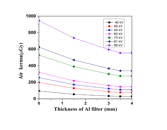
Analytic radiography is a normal image testing technique which has been utilized for quite a long time. It is recommended by specialists so they can identify any problem in patients' bodies without a cut. Thinking about its wide use, the principle objective of this investigation is to give a top notch picture by keeping the radiation portion as low as conceivable through identifying any variety in quality control (QC) boundaries. In this work, some standard quality control boundaries, for example, voltage exactness, time precision test, tube yield linearity, half value layer (HVL) of x-beam were measured. These quality control (QC) boundaries were estimated by a dosimeter keeping a distance of 100 cm from source. The voltage precision went from 0.31% to 4.67% and the time exactness test went from 0% to 2.29%. The consequences of this investigation show that all the QC boundaries are inside the acceptable level which guarantees the advancement of the low portion conveyed to the patients.
References
Abd-Alla, A., Salih, S. M., & Albashir, F. A. (2019).Evaluation of X- ray machines Gezira state: Based quality control. International Journal of Innovative Science, Engineering and Technology, 6(7), 68-71.
Abdulkadir, M. K. ( 2020) . Quality assurance in medical imaging: A review of challenges in Nigeria. Journal of Radiology Nursing, 39(3),238-244. doi:10.1016/j.jradnu.2020.02.005
Al- Kinani, A. T. , & Mohsen, Y. ( 2014) . Study of the quality assurance of conventional X- ray units at medical city in Baghdad. Arab Journal of Nuclear Sciences and Applications, 47( 2) ,129-137.
Asadinezhad, M., Bahreyni Toossi, M. T.,Ebrahiminia, A., & Giahi, M. (2017). Quality control assessment of conventional radiology devices in Iran. Iranian Journal of Medical Physics, 14(1), 1-7.doi:10.22038/ijmp.2017.19052.1173
Gholami, M., Nemati, F., & Karami, V. (2015). The evaluation of conventional X-ray exposure parameters including tube voltage and exposure time in private and governmental hospitals of Lorestan province, Iran. Iranian Journal of Medical Physics, 12(2), 85-92. doi:10.22038/ijmp.2015.4770
Health Canada. ( 2008) . Safety Code 35: Safety procedures for the installation, use and control of X-ray equipment in large medical radiological facilities. Health Canada.
Inkoom, S. , Schandorf, C. , Emi- Reynolds, G. , & Fletcher, J. J. ( 2011) . Quality assurance and quality control of equipment in diagnostic radiology practice-the Ghanaian experience. In I. Akyar (Ed.), Wide spectra of quality control (pp. 291-308). InTech. doi:10.5772/22591
International Atomic Energy Agency. (2007).Technical Reports Series No. 457: Dosimetry in diagnostic radiology: An international code of practice. Vienna, Austria: IAEA.
International Commission on Radiation Units and Measurements. (1993). ICRU Report 51,Quantities, quantities and units in radiation protection dosimetry. Bethesda, MD: ICRU.
Ismail, H. A., Ali, O. A., Omer, M. A, Garelnabi, M. E. , & Mustafa, N. S. ( 2015) . Evaluation of diagnostic radiology department in term of quality control (QC) of X-ray units at Khartoum state hospitals. International Journal of Science and Research, 4(1),1875-1878.
Rahman, M. S. , Kim, G. , Tsujimura, N. , Yoshida, T., Kanai, K., & Kurihara, O. (2008). Influence of the PMMA slab and ISO water phantom in calibrating personal dosimeter in the energy range of 36-662 keV. Journal of Nuclear Science and Technology, 45, 264-267. doi:10.1080/00223131.2008.10875838
Ranallo, F. N. (1998). Recommended standards for the routine performance testing of diagnostic X‐ ray imaging systems, IPEM Report No.77. Medical Physics, 25(8), 1556-1557.doi:10.1118/1.598335
Rubai, S. S. , Rahman, M. S. , Purohit, S. , Patwary, M. K. A., Meaze, A. K. M. M. H., & Mamun,A. A. (2018). Measurements of entrance surface dose and effective dose of patients in diagnostic radiography. Biomedical Journal of Science and Technical Research,12(1),1-5.doi:10.26717/BJSTR.2018.12.002186
Taha, T. M. (2011). Study the quality assurance of conventional X-ray machine using non-invasive KV meter. Proceedings of the Tenth Radiation Physics and Protection Conference (pp. 105-110). Cairo, Egypt.
Downloads
Published
How to Cite
Issue
Section
License
Copyright (c) 2023 Suan Sunandha Rajabhat University

This work is licensed under a Creative Commons Attribution 4.0 International License.